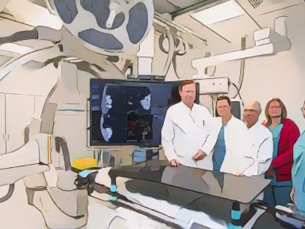
Modern technology takes center stage at MHH, as they revolutionize their stroke and cerebral hemorrhage treatments with a cutting-edge angiography system from Canon. This innovative device, which minimizes radiation exposure and offers exceptional image quality, has the potential to significantly improve patient outcomes.
Dr. Friedrich Götz, Head of Interventional Neuroradiology, explains the intriguing process. "We insert a telescopic catheter system into the affected cerebral vessel through the groin or forearm. By either sucking out or extracting the clot, we can clear the vessel for better blood flow," he says. The new device's precision and control elevate the procedure's success rate, leaving MHH's patients grinning with gratitude.
But that's not all. The system also boasts a VASO-CT feature, which excels at creating high-resolution cross-sectional images of small vessels. In the realm of aneurysms, this is invaluable, as the images allow physicians like Dr. Omar Abu-Fares to confirm if a stent is in full contact with the vessel wall or needs further adjustment.
It's essential to remember that this technology isn't just a novelty; it's a game-changer for Lower Saxony's medical schools and radiology departments. Dr. Heinrich Lanfermann, an MHH professor, sums it up: "This new device is a huge advantage for our patients, and it's an example of how we're constantly pushing boundaries to improve care."
Now, let's delve a little deeper into the inner workings of this remarkable device. At its core, the Canon Medical Systems' Adora DRFi features a fusion of static radiography and dynamic fluoroscopy. This unique combination allows for efficient workflows and optimal equipment utilization.
The system's high-resolution image detector shines when it comes to detailed imaging, which is indispensable for diagnosing and treating cerebral conditions with precision. Moreover, it likely employs advanced cone beam CT (CBCT) technology for high-resolution imaging, offering exceptional visual insight into constricted cerebral vessels.
Impressively, the Adora DRFi's VASO-CT can produce high-resolution cross-sectional images of even the tiniest vessels for unparalleled detail. This feature is key when assessing aneurysms, as it allows physicians to gauge stent placement and confirm full vessel wall contact.
The system's true power lies in its ability to acquire multiple X-ray projections during gantry rotation, back-projecting these images to form a volumetric dataset. This advanced technology allows physicians to visualize the angioarchitecture of cerebral diseases, aiding in more accurate diagnoses and informed treatment planning.
All these incredible features work together to provide high-quality images while minimizing radiation exposure, making the Adora DRFi a device that celebrates patient safety. Its efficiency in image acquisition and processing means quicker diagnoses and a more streamlined treatment process, further underlining the system's benefits.
In conclusion, MHH's incorporation of the Adora DRFi represents a major leap forward in neurovascular care. This remarkable device, with its advanced features and capabilities, combines precision, safety, and efficiency to support physicians in diagnosing and treating cerebral hemorrhage, stroke, and aneurysms with ease.


